In this article we are going to review the Basic Views of Trans esophageal Echocardiogram and the structures visible in each view. Watch the above video to view real time video clips of major TEE Windows with labeled structures in each view.
Long considered to be a vital diagnostic tool utilized exclusively within the realm of cardiology, transesophageal echocardiography (TEE) is being used safely and effectively within the peri-operative, critical care, and emergency room settings. Of particular importance is the ability to diagnose a vast array of diverse pathologies causing hemodynamic instability. In this capacity, a focused TEE examination has been proven to serve as an important adjunct to the history and physical examination to aid in the clinical decision-making process and to help guide treatment strategies.
TEE Vs TTE:
The transducers utilized in transthoracic echocardiography (TTE) are typically single-plane phased array probes, requiring the sonographer to physically manipulate the probe to move from one view to the next. On the other hand, most TEE probes, and for the purposes of the discussion in this chapter, are multiplane phased array probes. Several thousand piezoelectric crystal elements are assembled in a matrix configuration and mounted to the distal tip of an endoscope. This arrangement allows a linear scan line to be created that can be electronically rotated from 0 toward 180 degrees using two buttons on the handle of the ultrasound probe as shown in the figures in the above video.
Transesophageal echo probe typically has a large control wheel, a small control wheel, and knobs to adjust the angle of the ultrasound beam.
TEE Probe Manipulations:
Physical probe manipulation can be performed by turning the large control wheel on the handle to anteflex or retroflex the probe. Furthermore, the tip of the probe can be flexed left or right by rotating the smaller control wheel. The shaft of the probe can be advanced or withdrawn. it can also be turned clockwise to image right-sided structures, or turned counterclockwise to image left-sided structures
As such, multiple views are able to be created without any physical probe manipulation. Rotating the scan line (also called the omniplane angle ) in the direction of 180 degrees is termed rotating forward , whereas rotating back to 0 degrees is termed rotating backward .
By these manipulations we can get Views at Mid esophageal level which is at the distance of about 30 to 40 cm from incisor teeth, upper esophageal level 20 to 25 cm. and Trans-gastric views are obtained at 40 to 50 cm.
Let’s review each view and the major structures visible in each view.
Mid Esophageal Four Chamber View
For Mid Esophageal four chamber view, probe should be advanced into the esophagus approximately 30 to 40 cm. The angle should be set to between 0 and 20 degrees. In the adult chest, the imaging depth should initially be set to about 16 cm to ensure the entirety of the heart is visualized. If the aortic valve and left ventricular outflow tract are still visible (informally called a five-chamber view ), the probe should be advanced slightly into the esophagus until the LVOT disappears, and only the four chambers are visible, and the annulus of the tricuspid valve is maximized. these are the structures visible in this view, we can appreciate Left atrium, right atrium, Left ventricle and right ventricle.
Now apply color Doppler and take necessary measurements if anything abnormal is found.
Mid Esophageal Four Chamber View with LVOT
By withdrawing probe a little we can get Left outflow tract into view to make it a four chamber view with Left outflow tract, in other words it’s a five chamber view. Apply color Doppler on outflow tract and take necessary measurements.
Mid Esophageal Bi commissural View
Mid Esophageal Bi commissural View is obtained by angling the beam to about 60 to 70 degrees, it’s a great view to assess mitral valve scallops. Probe can be directed anterior or posteriorly to assess all the scallops. In the mid-way between anterior and posterior P1 , A2 and P3 scallop can be visualized.
Mid Esophageal Two Chamber View
With only minor physical manipulation of the probe, the omniplane angle should be rotated forward to between 80 and 100 degrees. The left atrium (top) and left ventricle (bottom) will both be visible. The anterior and inferior walls of the left ventricle will appear on the right and left of the screen, respectively. The left atrial appendage will be visible and can be assessed for clots.
Mid Esophageal Long Axis View
The omniplane angle is further rotated from the Mid Esophageal two chamber view to between 120 and 160 degrees. Again, both the left atrium and left ventricle are visible. The mitral valve resides on the left side of the screen, and the long axis of the aortic valve can be visualized on the right side of the screen at the distal end of the LVOT as it opens to the proximal ascending aorta. The anteroseptal wall separates the left ventricle from the right ventricle outflow tract. The inferolateral wall of the left ventricle is visible on the left side of the screen.
Mid Esophageal Bicaval
From the mid esophageal long axis view, the shaft of the probe should be turned clockwise and the omniplane angle rotated backward to between 80 and 110 degrees to get this View. The depth should be decreased to maximize visualization of all the relevant structures.
The inter-atrial septum can be visualized, separating the left atrium from the right atrium. Assessments for the presence of a PFO, or inter-atrial shunt should be made utilizing color flow Doppler.
Mid esophageal RV Inflow /Outflow view
Without moving the probe, the omniplane angle should be decreased even farther to approximately 50 and 90 degrees. The valve on the left is the tricuspid valve as blood inflows into the right ventricle. Toward the right of the screen, the long axis of the pulmonic valve appears and serves as the outflow of blood from the right ventricle, hence the name of this view.
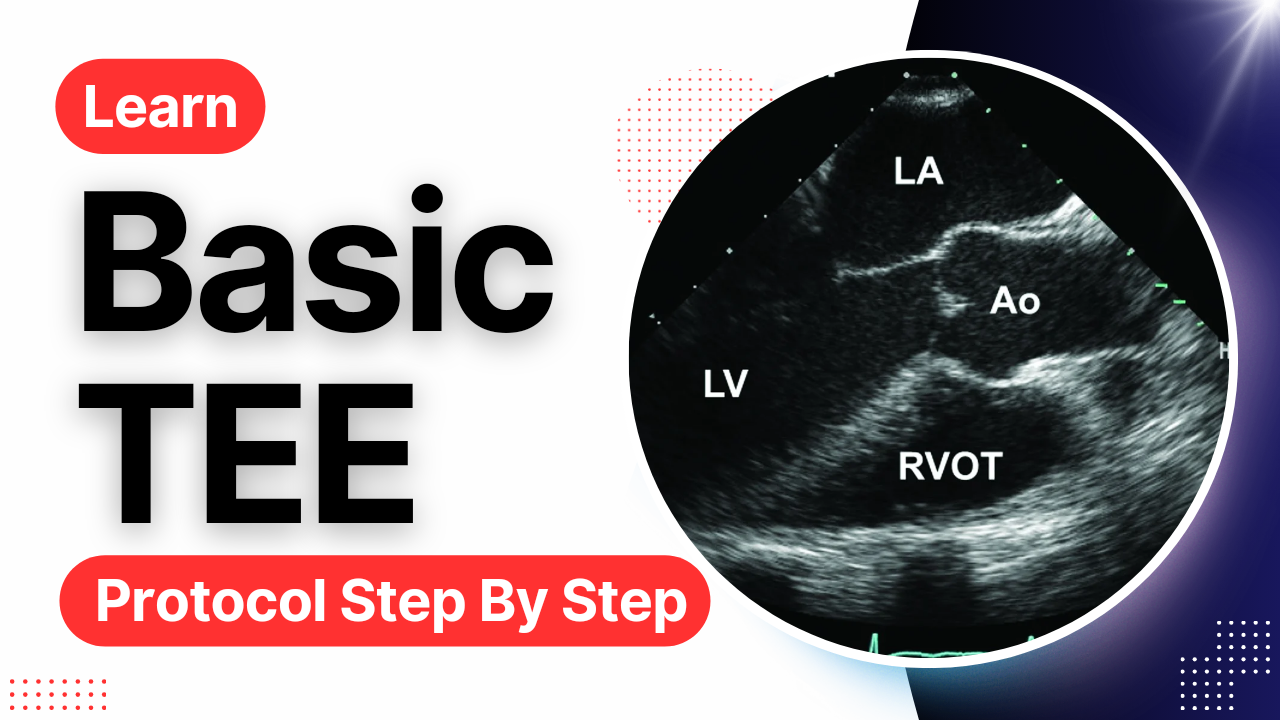
Mid esophageal AV SAX
Decreasing the omniplane angle from the RV inflow–outflow view to between 30 and 60 degrees will help to visualize all three leaflets of the AV in short axis. Typically, this involves only a slight decrease in the omniplane angle, if at all.
Ascending Aorta LAX
To get this view adjust angle 100 to 150 degrees, its best obtained from mid esophageal Long Axis View , by slowly withdrawing the probe while keeping aorta in view by making small clockwise and counterclockwise adjustments.
Descending Aorta SAX
Withdraw the probe back to the mid esophageal four chamber view and rotate the shaft of the probe to 180 degrees until the descending aorta comes into view. The depth should be decreased to approximately 6 to 8 cm to maximize the size of the aorta. The probe should be advanced and withdrawn to scan as much of the descending aorta as possible starting from the proximal descending arch. The short axis of the aorta is visualized and should appear circular.
TG
Standard Trans-gastric views are obtained by advancing the probe into the stomach at about 40 to 45 cm from the incisor teeth. At zero degrees Short Axis View is obtained, showing the Left ventricular walls at papillary muscle level. Note that by looking from the stomach, first wall be inferior, so that is on the top of the screen, and anterior wall is at the bottom of the screen.
By manipulating the probe to basal level, we can get short axis view of mitral valve.
By adjusting transducer imaging plane angle we can obtain cuts through the long axis of the heart, at about 90 degrees, two chamber view is visualized.
Trans-gastric long axis view is obtained By further adjusting the angle to about 100 to 120 degrees .
For Right ventricle Inflow view adjust the angle to more than 120 degrees, and slightly ante flex the probe.
By advancing the probe further deep to about 50 cm we get a deep trans-gastric long axis view. Here Left ventricle is foreshortened but this view is great to assess left ventricular outflow tract and its gradients.
Here is the summary of all the major views we covered. We can get more views by little manipulations according to the pathology found.
Key Views of Transesophageal Echocardiogram:
Mid Esophageal 30 – 40 cm
Four Chamber View
Four Chambers with LVOT
Mitral Commisural View
Two Chamber
Long Axis
Bi Caval
RV Inflow- Outflow View
AV SAX
Ascending Aorta
Descending Aorta
Upper Esophageal 20 – 25 cm
Aortic Arch SAX
Aortic Arch Long Axis
Transgastric Views 40 – 50 cm
Mid Short Axis
Basal Short Axis
Two Chamber view
Long Axis View
RV Inflow View
Deep Transgastric Long Axis
If you have any queries let me know in the comments section below , and to practice Transesophageal Echocardiography cases, Visit our channel:
https://www.youtube.com/@cardionotes
Thanks.